how do they x ray babies hips
At birth the baby cant move the thigh outward at the hip as far as normally possible. Babies and toddlers dont need teeth to eat successfully.

Infant Diagnosis International Hip Dysplasia Institute
An ultrasound may be needed to get a picture of the hip.

. Your doctor may ask for an ultrasound or X-ray of the hip joint to diagnose DDH. Appointments and Referrals. These tests expose children to low doses of radiation.
1317 years average 15 years. Place the infant supine with the hip flexed to 90 in a neutral position. Hip ultrasounds take less than 20 minutes and the child will not feel any pain during the examination.
What Are the. Computed bone maturity bone age assessment. The age groups were based on exposures suitable for tissue thickness in the direction of the X-ray beam of a patient of averagestandard size in that age group for each projection.
You will go in the room with him he will need to be stripped from the waist down they will take x-rays of him flat on his back legs dead straight and together you wil be able to hold him in this position then an x-ray of his still on his back with his knees bent facing outwards and the soles of his feet put together he will be fine. It is put on by an orthopedic surgeon while using x-ray to make sure the hip is aligned correctly. Inflammation where your sacrum joins.
Pregnancy usually occurs by sexual intercourse but can also occur through assisted reproductive technology procedures. Im a nurse in a pedi operating room and Ive done them many times The hip dysplasia usually resolves on its own with the casting or bracing as the baby gets older. After around 4 to 6 months of age X-rays are the preferred method for evaluating and monitoring hip dysplasia.
X-rays have more energy than rays of visible light or radio waves. Because of the risk of developmental dysplasia of the hip in infants born breech-despite a normal physical exam-the American Academy of Pediatrics AAP guidelines recommend ultrasound US hip imaging at 6 weeks of age for breech females and optional imaging for breech males. They do this by gently pushing and pulling the babys thigh bones to see if they are loose in the hip socket.
After around 4 to 6 months of age X-rays are the preferred method for evaluating and monitoring hip dysplasia. Two tests are performed called the Barlow and Ortolani tests to examine the function of the hip joints. Perthes disease also known as Legg-Calvé-Perthes disease is an idiopathic avascular necrosis of the proximal femoral epiphysis.
X-rays can be taken once your baby is 3 months old. Ultrasounds use inaudible sound waves which bounce off of the bones and muscles to create an image for radiologists to interpret. The doctor hears or feels a hip click when moving the infants thigh outward.
They can penetrate your body. Because they spin around the body taking multiple images CT scans can deliver radiation doses that are up to 200 times higher than an. This test attempts to detect joint subluxation or dislocation by trying to displace the femoral head posteriolaterally from the acetabulum.
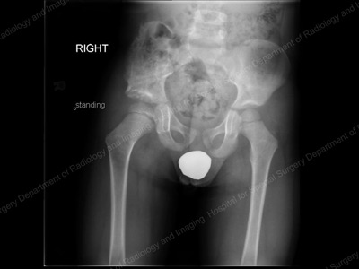
The doctor first checks your babys hips in the hospital after birth. X-rays can be taken once your baby is 3 months old. A hip ultrasound might be done for a baby if the doctor finds a hip problem such as.
Babies with DDH can be successfully treated with a special brace. About one in eight scans ordered for kids is a CT scan. How is hip dysplasia treated in babies.
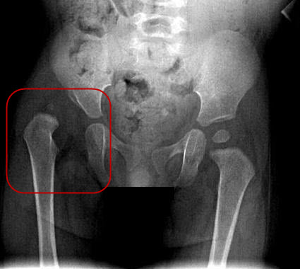
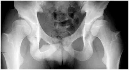
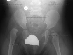
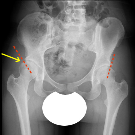
This x-ray shows a dislocated hip on the patients right side. The purpose of this study is to report US results and follow-up of. Its a cast that goes around both hips and down the leg to keep the hips aligned.
Hip problems may not be present at birth. 37 years average 5 years. Most children do not need surgery but for those who do an arthrogram x-ray dye injected into the hip joint at the beginning of the surgery can help the surgeon decide exactly what needs to be corrected.
Arthritis that affects your hip. They are of more concern. X-rays are forms of radiant energy like light or radio waves.
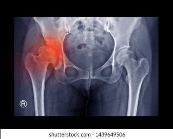
A hip click can be felt by the examiner when the hip joints may not have formed normally. During treatment x-rays can reveal the progress of the hip as it improves. Hip ultrasounds are a safe non-invasive procedure that does not use any radiation.
However x-rays of the mothers lower torso - abdomen stomach pelvis lower back or kidneys - may expose the unborn child to the direct x-ray beam. It occurs more commonly in boys typically between 5 and 8 years of age but may range from the ages 3-12. It could even be as simple as a urinary tract infection.
In babies with hip dysplasia the joint has not formed normally and the hips are prone to moving in and out of joint. A pelvic X-ray can help your doctor detect various conditions such as. Hip ultrasounds are a safe non-invasive procedure that does not use any radiation.
Its a cast that goes around both hips and down the leg to keep the hips aligned. Computed bone maturity bone age measurement are performed in cases of suspected growth delay or early pubertal development. They give your healthcare provider information about structures inside the body.
Place your index and middle finger along the greater trochanter and your thumb on the inner thigh. 812 years average 10 years. During the examination they examine the X-rays that the technicians take of their patients teeth and perform a full examination of the teeth gums tongue and jaws to find out if there are any problems.
But for babies with an abnormal physical exam or major risk factors for developmental dysplasia of the hip or DDH family history Breech position etc the AAP supports referral for. Computed tomography scanogram for leg length discrepancy assessment. The American Academy of Pediatrics does not recommend routine ultrasounds for every infant.
Then a surgeon gently pushes the ball of their thighbone joint into the hip socket where it belongs. It can occur bilaterally but it is usually asymmetric. If a physical exam an ultrasound or an X-ray confirm a diagnosis your pediatrician will likely refer you to a pediatric orthopedic specialist for continued care and treatment.
X-rays are a kind of imaging test.

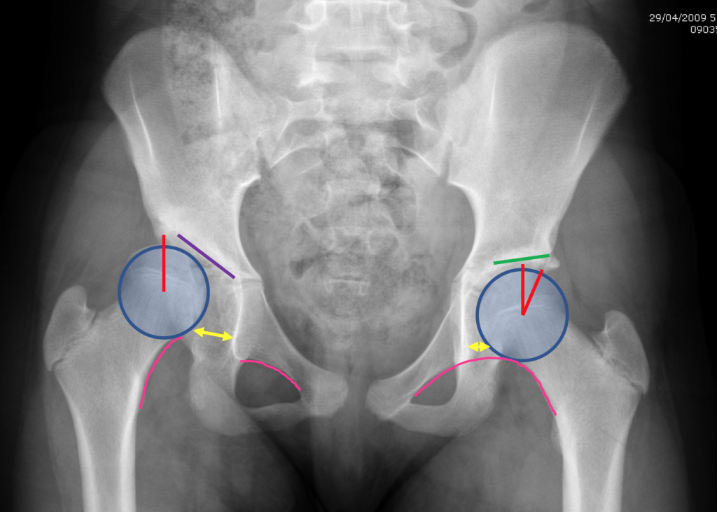
Hip Dysplasia What S With All The Angles
Developmental Hip Dysplasia In Babies And Young Children
Developmental Hip Dysplasia In Babies And Young Children
Pediatric Hip Disorders Radsource

Hip Dysplasia Should My Child Be Screened Uva Radiology
X Ray Screening International Hip Dysplasia Institute

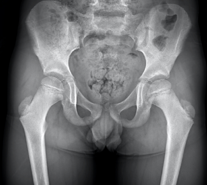
Normal Pelvis X Ray 4 Year Old Radiology Case Radiopaedia Org

Pelvis X Ray Ap View Showing Left Sided Dysplastic Hip With Femur Download Scientific Diagram
Hip Dysplasia Information Symptoms Diagnosis Treatment

Infant Diagnosis International Hip Dysplasia Institute
Infant Diagnosis International Hip Dysplasia Institute
Developmental Hip Dysplasia In Babies And Young Children

Pediatric Hip Frog Leg Lateral View Radiology Reference Article Radiopaedia Org
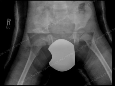
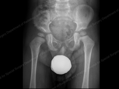
If You Ve Ever Wondered How They X Ray Babies R Damnthatsinteresting
Hip Dysplasia Adolescent Description
Congenital Hip Dysplasia Symptoms Treatments Orthopedics
Developmental Hip Dysplasia In Babies And Young Children

Developmental Dysplasia Of The Hip Radiology Reference Article Radiopaedia Org
Hip Dysplasia Images Stock Photos Vectors Shutterstock